How reliable are X-rays?
How reliable are x-rays? They are only as trustworthy as the reporter and what is actually to be seen.
Daniel D. Palmer founded what we today know as chiropractic; coincidentally it was in the same year, 1895, that Wilhelm Röntgen discovered X radiation.
Ever since, radiography and Chiropractic have had much in common in the treatment and diagnosis mainly of bone and joint conditions.
But are they trustworthy? We can consider this under these four subheadings.
- How well trained are General Practitioners and Chiropractors in the reading of X-rays?
- How trustworthy are the radiologist's findings?
- Is the suspected lesion adequately visualised on the radiograph?
- Could the identified faulty structure be an incidental finding? True, but quite unrelated to the patient's complaint.
Ionising radiation
How safe is ionising radiation? Not at all unfortunately is the best answer. Early physicists and radiological technologists suffered from a much higher rate of breast and skin cancer; and leukemia. Once they realised the danger they were in, protective clothing and lead shielding was used. Now it's the ailing public who are at risk.
Chiropractors have taken a leading role in the development of X-ray technology and one of the best textbooks is written by two amazing DCs.
Essentials of skeletal radiology is a standard textbook today.

Every chiropractor goes through extensive training, not only in the taking of X-ray, but in reading them.
Whilst there have been huge advances in radiological technology, reducing damaging exposure, research in medical journal Lancet reports that at least seven percent of cancer today is caused by ionising radiation from man-made sources. Mainly from X-rays, CT scans and mammograms.
To the extent that every doctor has to ask the question: do I really need to expose this patient to X-rays?
We all look forward to the day when MRI becomes more cost effective, and X-rays and CT scans can be greatly reduced.
A CT delivers a similar amount of radiation as the Hiroshima atomic bomb[3].
The now clear-cut evidence that we doctors have actually caused many of our patients' cancers, known as iatrogenic illness has led to another complication.
Both doctors and radiologists are increasingly reluctant to order X-rays, or request only limited views, increasing the risk of missing an important diagnosis.
How reliable are X-rays?
How reliable are x-rays? Scans are better but CT might make you glow in the dark. MRI has the advantage that it is not ionising radiation.
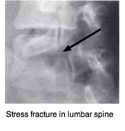
A thirteen-year old adolescent was knocked over with resulting acute low back pain. It was important to rule out a fracture, particularly through a narrow isthmus of bone resulting in a "spondylolysis" as an adjustment would obviously be contraindicated.
In fact it would certainly aggravate the condition, possibly resulting in a spondylolysthesis.
The
medical radiologist refused to take the necessary extra X-rays, citing
the danger of excessive radiation. Perhaps correctly so, but we missed a
stress fracture through the "pars interarticularis", not seen on 20% of
plain film without "obliques."
Research by Katoh, Ikata and Fujii reveals there is a likelihood of healing of the fracture with early diagnosis and immobilisation, but late stage lesions rarely regrow. NSAIDS are contraindicated according to Syrmou et al (Hippokratia 2010 Jan) because they "slow down bone growth and healing."
Spondylolysthesis
Spondylolysthesis occurs when this pars fracture occurs bilaterally; but in answer to our question, how reliable are x-rays, it's often missed.
Case 2.
A forty year-old man fell about three metres, with immediate severe pain in the right foot. Within a short period the lower leg was oedematous and throbbing. He was unable to stand on the leg.
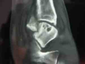
X-rays of the ankle were declared normal. Here they are.
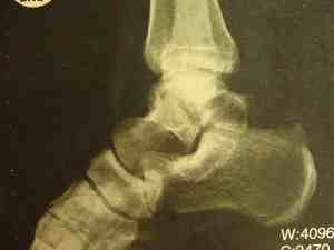
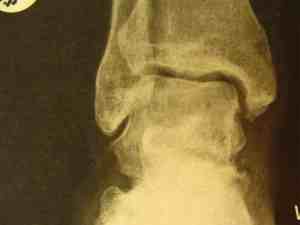
Normal X-rays?
He was diagnosed with a bad sprain of the ankle, and put into a boot to limit movement.
He first consulted me a year and a half later. The ankle was still swollen, and there was considerable discolouration of the skin. Eversion was severely limited.
Fractures in the ankle are notoriously difficult to see because of the overlapping structures. A scan revealed the partially healed fracture through the talus bone.
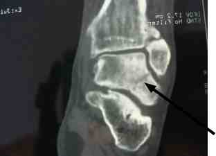
Worse, because of the disrupted blood supply to the talus, the main bone joining the leg to the foot is dying. A process called "avascular necrosis."
Can you see the great holes of dead bone in the talus? The fracture cut off the blood supply to the osteoblasts. It should have been pinned immediately.
How reliable are X-rays? Sometimes fractures can only be seen on a scan.
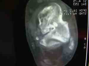
Whilst gentle Chiropractic mobilisation of the joints around the talus brought about 50% relief of pain and stiffness for about six months, it's proved temporary. The talus is a major weight-bearing bone. A total ankle replacement is on the cards.
Not because of the original injury, but because the missed fracture led to inappropriate treatment.

Case 3.
Radiologists are totally dependent on the doctor for supplying relevant clinical information.
A young man had a bizarre accident in which a very heavy concrete pillar fell on him, forcing his spine into flexion. He had immediate severe rib (and back) pain. His doctor suspected a rib fracture (he was right) and sent him for rib x-rays. No mention was made of possible spinal fractures.
The radiologist commented only on the ribs. Whether he missed these compression fractures, or only commented on what was asked by the doctor, is immaterial. The ribs have healed, but the young man continues to have severe on-going midback pain some three years after the trauma.
How Reliable are X-rays? Not as reliable as a good clinical examination.
Case 4.
A middle aged man began to experience lower neck, midback pain and tingling in the left arm.
His doctor ordered X-rays. Here's the initial report:
Report I:
Normal vertebral body alignment. No traumatic fracture or dislocation. The intervertebral disc spaces are preserved at all levels with no spondylotic or disc degenerative changes. No subluxation of the verebral bodies. Normal alignment of the spinous processes and facet joints. On the oblique views, the bony neural exist foramina are patent at all levels. The precervical soft tissue is normal.
Radiologist's comment:
- No features of trauma.
- No spondylotic changes.
Based on the radiologist's report his doctor said there was no need for concern and sent him for physiotherapy.
Aside: Worldwide medical doctors have little training in reading of X-rays so they are reliant on the radiologist's report.
Three months later he first consulted me with serious "hard" neurological findings. Not satisfied with the original report, I asked for a review of the X-rays by another radiologist. Here's his report:
Report II:
Bony alignment is normal. Congenital ankylosis between C2 and C3. Disc spaces are preserved, although the C7-T1 disc is not well shown on the lateral view. Quite large degenerate spurs encroach on both C6-C7 foramina. These arise from the uncovertebral joints. If superadded disc protrusion is suspected an MRI scan may be of help. No cervical ribs.
Notice that the first radiologist missed two important facts:
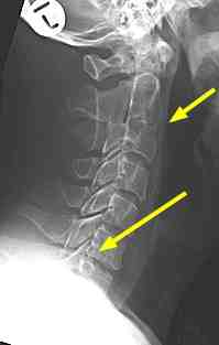
- He was born with an anomaly: C2 and C3 have formed one "block" vertebra.
- Quite advanced spondolytic changes at C6-C7.
Tingling in arms and hands
How reliable are x-rays in diagnosing the source of tingling in arms and hands?
Tingling in arms and hands usually comes from degenerative change in the lower cervical spine. Very rarely it occurs because of anatomical changes that occur as the spinal cord descends through the foramen magnum at the base of the skull.
But disc prolapse is poorly assessed on plain film.

- Tingling in arms and hands is often the result of degenerative changes emanating from small joints located only in the lower neck. Known by various names today they are mostly called the UncoVertebral Joints or the the Joints of Luschka.
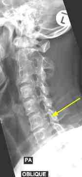
The left oblique view below show the large degenerate spur emerging from the UncoVertebral joint Luschka ... see how it's invading the foramen. The nerve root takes up about half the space in the IVF (inter vertebral foramen) so any tissue that is "space-occupying" will threaten the nerve root.
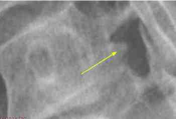
The right foramen shows how these degenerative changes are occurring in the opposite side also.

If the nerve root is frankly pinched then it causes a deep, extremely
painful ache in the arm. Turning the head to that side, and
simultaneously looking up causes immediate pain or tingling in the arm.
The dermatomal pattern can be extremely useful in localising the lesion. Take particularly note of which part is affected.
Nerve roots supply a particular, defined part of the skin. So it's not just generalised tingling and numbness in the arm and hand. In this case, the nerve passing through the foramen beween C6-C7 is the C7 nerve root. It supplies the middle finger, and possibly the adjacent index and ring fingers.
You'll notice that the dermatomal pattern was again the clue in Case 5.

Case 5
X-rays show primarily bone, though soft tissue densities like fluids and gases can be seen to some extent.
But an X-ray gives little indication of the extent of a recent disc injury, being soft tissue. Old disc injuries result in loss of disc space, but a new injury may be totally unvisualised on plain film.
In this case a 65-year old chiropractor (me!) bent, twisted and pulled a very heavy patient into position in preparation for a sacro-iliac joint manipulation.
You can read the details of the case if interested, at Femoral nerve damage but for the purposes of this page let us compare the lateral lumbar as seen on X-ray and on MR.
The patient had a long history of lower back pain episodes (which gardeners, glider pilots, beekeepers don't?) but Chiropractic treatment of the L4-L5 and sometimes L5 on S1 joints soon rectified the subluxation.
But this time, after a stupidity, the pain radiated down the side and front of the thigh to the knee. The Femoral nerve, so his chiropractor (my daughter!) was fairly sure the problem was higher.
Here is the X-ray.
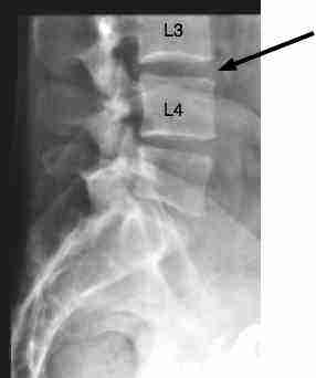
Really all three visualised disc spaces look pretty good, with L3-L4 the best of them all; and there's where her fingers were telling her the problem was.
Because of the severity of the leg pain, the progressive numbness in the lower limb just below the knee and weakness developing in the quadriceps muscles, we jointly agreed an MR was necessary.
The absence of lower back pain was an ominous sign, incidentally.
Femoral nerve damage
How reliable are x-rays in assessing femoral nerve damage?
Femoral nerve damage often causes the knee to collapse due to a weak quadriceps muscle.
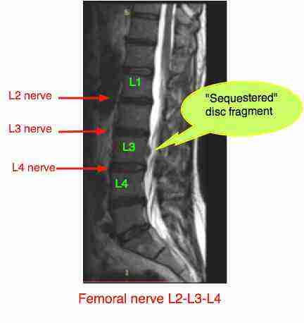
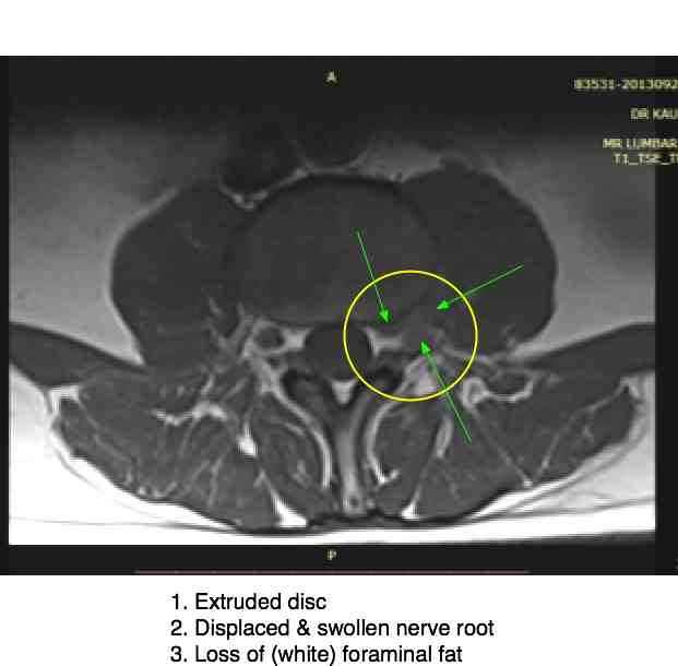
Oh dear, a Chiropractor's worst nightmare, a sequestered disc fragment lying in the spinal canal. But did it come from the L2-L3 disc or L3 to L4 disc? This is normally considered a surgical emergency.
The MR of the L3-L4 disc (remember, the best looking one on the radiograph) shows a severely extruded disc pinching the L4 nerve root. Hence the anterior leg pain.
To the surgeon? Read on at Femoral nerve damage ...
How reliable are X-rays? Sometimes not at all reliable. The radiologist's report states (with which I agree): "Minimal loss of disc height is present at L4-L5 and L5 to S1."
There was no mention whatsoever of L3-L4.
Anyone relying entirely in the X-rays report would have been led entirely astray. This man is shirking!
No sirree, this man is not shirking, he was in severe pain. X-rays are not good for determining the level and severity of a disc injury.
How reliable are X-rays?
How long is a piece of string? The point of this letter is not to point fingers at others who have missed serious diagnoses. All doctors screw up periodically, myself included.
"Sue the bastards," might be the reaction of some. In my opinion there's a very fine line between an honest mistake and negligence worthy of being taken to court for damages.
Every doctor makes mistakes and, if you going to sue him every time, all that happens is that malpractice insurance premiums soar, which the patient pays for ultimately; and you end up with totally stressed, exhausted health professionals who have to cover their butts at every turn, running up yet more expensive tests.
Expenses which increasingly the poor and middle-class folk can ill afford.
They are not only expensive, but also dangerous tests. To be absolutely sure you don't have a fracture, even when clinically the likelihood is small, do you really want your doctor to expose you to so much radiation that you glow in the dark?
What's the point? There is no perfectly reliable test. The responsibility is on both the patient and doctor, in an honest relationship, to evaluate progress. If you know you are not getting better and there is definitely something wrong, for heaven's sake, make it abundantly clear to your chiropractor, medical doctor and dentist.
If your doctor won't listen, go elsewhere for a second opinion.
A thorough examination
How reliable are X-rays?
Much of these problems could have been avoided had the patient been carefully and thoroughly examined. And if we took the time to respect and listen to each other.
In case I, had the radiologist just listened to me, the attending doctor it would have been different. I sent the child back for a second time, only to have the tests refused a second time.
In the second, the patient was continually berated for putting it on. "There's clearly nothing wrong, get on with your work." Had the relevant health professionals just listened properly to the account of the suffering, things would have worked out differently.
In the third simple percussion over the spine would have revealed not only rib injuries, but the likelihood of a spinal fracture.
And lastly, in the fourth simple testing of a reflex and the strength of the muscles in the arm would have indicated the need for more tests.
Useful links
When browsing these links use right click and "Open Link in New Tab", or you may get a bad gateway signal.
- Home
- Femoral nerve
- How Reliable Are X-rays
Did you find this page useful? Then perhaps forward it to a suffering friend. Better still, Tweet or Face Book it.
